Shop No. 2-B, Opposite Arya Girls College, Shahabad, 136135, India


Best Study Visa Consultants in Shahbad Markanda
Dreamsky focus is on the individual from appreciating self-expression to encouraging original thought. And from fostering personal achievement to ensuring Long-term success. The road to your future begins here at Dreamsky. We’ll help you reach beyond all the barriers. That helps you to discover yourself and help you to become the kind of individual, you always knew you could be! Moreover, you have unique opinions and original thoughts. Along with this, you also have goals and Dreams, and your own ideas on how to make them come true. Your definition of “success” is unlike anyone else’s. And that’s what makes Dreamsky . For you!

REALIZE DREAMS
Dreamsky is one stop solution for all your study Abroad, IELTS, PTE, Spoken English, Interview Preparation, Personality Development .The core activity lies in assisting students to make the right choice with regard to pursuing education in overseas educational institutions.

GUIDANCE
Our expert consultants can hold the students hand through the entire process. As well, help them in choosing the right course befitting their interests and aptitude. Along with this, the entire process of course selection will be simplified. As we furnish all the vital information for choosing the correct study program.

STUDY ABROAD
In many countries like the USA, UK, Canada, Australia, New Zealand etc… In these countries, there are numerous options for further studies. And at Dreamsky we strive for success for our students.
STUDY DESTINATION

STUDY iN CANADA

CANADA

Canada is prominent for its education programs offering various streams to study in Canada. Come and Enroll.

STUDY iN NEWZEALAND

NEWZEALAND
New Zealand offers diverse range of options and strong international rankings to study in New Zealand.

STUDY iN U.S.A

U.S.A
USA is long been the international students favored destinations and to study in USA you have an..
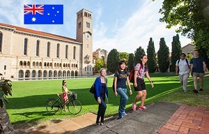
STUDY iN Australia

Australia
Australia offers a range of courses and the qualification framework to study in Australia is a quality assured…
Important Facts
Students Visa
IELTS
PTE
Spoken English
STUDY DESTINATION

SPOKEN ENGLISH
English is a universal language. As it is widely used all around the world. It unites people from all parts of the world. And the reason behind this, as it is an important means of communication.
Read more
INTERVIEW PREPARATION
We help students who are looking for job or admission in college by guiding them for their interview. We help students to crack their interviews effectively in affable manner.
Read more
PERSONALITY DEVELOPMENT
Personality development by name means to develop personality of a person. There are many courses that demand you to develop your personality. What is personality development?
Read moreClient Feedback
MR. AJAYDEEP SINGH
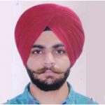
I am very thankful to Dreamsky regarding filing my Study Visa for Canada. The VISA results of Dreamsky are surely 100%. The staff of Dreamsky is very friendly.
MR. PARAS NARANG
They guided me very well, helped me at every stage while selecting the college and giving detailed information on the file preparation. Thanks a lot Dreamsky
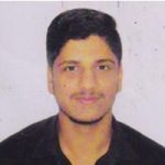
MR. ABHISHEK

The VISA results of Dreamsky are surely 100%. The staff of Dreamsky is very friendly.
MR. AJAY
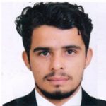
When I decided to go abroad for higher education, I was looking for right guidance and when I met counselor of Dreamsky my all worries were gone. I feel they made my dream come true with their sincere effort. Thanks for everything.